Myosin at work
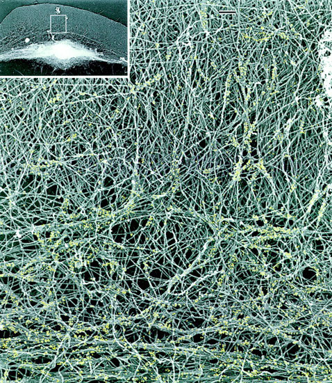
Myosin (gold dots) pulling together the actin filaments (grey) as seen under an electron microscope. The cell is moving towards the top of the image (see inset for a lower magnification image). Towards the bottom of the image, the increasing amount of myosin is pulling on the loose mesh of actin and collapsing the actin onto itself. The mesh is attached to the front of the cell so it compresses forwards, dragging the rest of the cell with it.
Figure courtesy of Gary Borisy. Copyright The Rockefeller University Press.